


When analyzing particulate materials, understanding particle size distribution is essential for predicting performance in applications ranging from pharmaceuticals to industrial powders and environmental science. There are two dominant paradigms for sizing particles: counting individuals or 動的画像解析 measuring their volumetric weight, and each provides distinct insights that can lead to very different conclusions. Imaging technologies have become indispensable tools in revealing the true nature of these distributions, offering visual and quantitative data that traditional sizing techniques often miss.
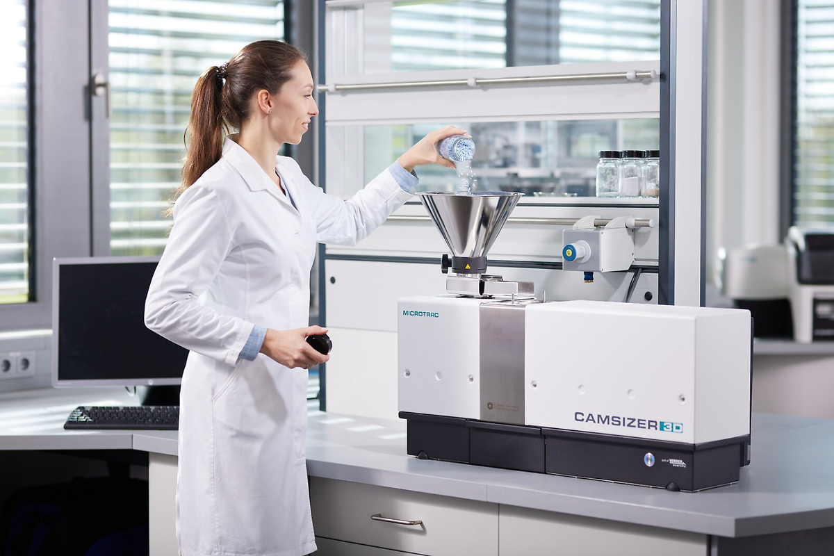
Measuring particle size by number means counting individual particles and determining how many fall into each size class. When the quantity of particles is more relevant than their bulk, number-based sizing is preferred,—for example, in aerosol science where inhalation exposure depends on particle count, or in nanomaterials where biological interactions are often governed by surface area and concentration of individual entities. Digital imaging tools allow precise counting of particles at the micro- and nanoscale, providing a clear picture of how many particles exist at each size. The number-based approach brings attention to micron-scale or nano-scale entities that dominate exposure or reactivity, leading to more accurate risk and efficacy assessments.
In contrast, measuring by volume assigns weight to each particle based on its three-dimensional size, skewing results toward the most voluminous individuals. A single large particle can dominate a volume-based distribution, even if thousands of smaller particles are present. This is often the preferred method in industries where flow properties, settling rates, or mixing behavior are critical—such as in concrete production or paint formulation. Volume-based methods like laser diffraction are common in these fields because they correlate well with bulk material behavior. However, they can obscure the presence of small particles that contribute little to volume but may significantly influence other properties.
Imaging bridges the gap between these two methods by allowing direct visualization of particle morphology and size. In contrast to models based on averaged physical interactions, imaging reveals irregular shapes, agglomerations, and surface features that profoundly affect how particles behave. What looks like a single large sphere may be a fused assembly of nanoparticles, leading to misinterpretation of its true nature. The source of volumetric dominance can be clarified: isolated giants or aggregated clusters.
Moreover, imaging enables the calculation of both number and volume distributions from the same dataset. Automated image analysis software calculates volume for each detected particle using its spatial dimensions, and then generate corresponding number and volume distributions side by side. Comparing both distributions side-by-side highlights anomalies undetectable by conventional tools. For instance, A bulk volume profile may appear homogeneous, while particle counts reveal a bimodal population. This indicates potential instability or contamination.
The practical implications of this distinction are significant. Drug formulations might pass bulk size criteria, yet imaging exposes an excess of nanoparticles that bypass therapeutic targets, potentially leading to wasted dosage or unintended systemic absorption. In environmental monitoring, a volume-based measurement of airborne dust might suggest low risk, but imaging could expose high concentrations of ultrafine particles capable of penetrating deep into the respiratory system.
Ultimately, imaging transforms particle size analysis from a statistical exercise into a visual science. It compels a shift from mathematical estimations to direct physical observation. The number perspective highlights individual risk; the volume perspective captures systemic effect. A triad of data: count, volume, and image, delivers the full story of a particulate material. Depending solely on volume or number leads to blind spots; imaging-integrated analysis eliminates ambiguity.